38 brain mri with labels
Labeled imaging anatomy cases | Radiology Reference Article ... This article lists a series of labeled imaging anatomy cases by body region and modality. Brain CT head: non-contrast axial CT head: non-contrast coronal CT head: non-contrast sagittal CT head: angiogram axial CT head: angiogram coronal CT... › pmc › articlesA novel biomarker of amnestic MCI based on dynamic cross ... Oct 20, 2015 · Conspicuous brain abnormalities that could account for cognitive decline were excluded using structural magnetic resonance imaging (MRI) data. The baseline neuropsychological evaluation covered the following cognitive domains: episodic and working memory, attention/psychomotor processing speed, executive function, language, and visual ...
101 labeled brain images and a consistent human cortical ... - PubMed given how difficult it is to label brains, the mindboggle-101 dataset is intended to serve as brain atlases for use in labeling other brains, as a normative dataset to establish morphometric variation in a healthy population for comparison against clinical populations, and contribute to the development, training, testing, and evaluation of …

Brain mri with labels
IMAIOS IMAIOS Brain MRI Atlas on the App Store Brain MRI Atlas is a FREE app that allows you to navigate through hundreds of of labeled brain structures. It is designed for all healthcare professionals as an interactive study and reference tool. Program Features: - Serial sequential axial T2 FLAIR images of the brain. - Structure labels organized by category. MRI anatomy | free MRI axial brain anatomy - Mrimaster.com This MRI brain cross sectional anatomy tool is absolutely free to use. Use the mouse scroll wheel to move the images up and down alternatively use the tiny arrows (>>) on both side of the image to move the images.
Brain mri with labels. Brain MRI segmentation | Kaggle Journal of Neuro-Oncology, 2017. This dataset contains brain MR images together with manual FLAIR abnormality segmentation masks. The images were obtained from The Cancer Imaging Archive (TCIA). They correspond to 110 patients included in The Cancer Genome Atlas (TCGA) lower-grade glioma collection with at least fluid-attenuated inversion ... Arterial spin labeling MRI: clinical applications in the brain Visualization of cerebral blood flow (CBF) has become an important part of neuroimaging for a wide range of diseases. Arterial spin labeling (ASL) perfusion magnetic resonance imaging (MRI) sequences are increasingly being used to provide MR-based CBF quantification without the need for contrast administration, and can be obtained in conjunction with a structural MRI study. Atlas of BRAIN MRI - W-Radiology Brain magnetic resonance imaging (MRI) is a common medical imaging method that allows clinicians to examine the brain's anatomy (1). It uses a magnetic field and radio waves to produce detailed images of the brain and the brainstem to detect various conditions (2). Brain lobes - annotated MRI | Radiology Case | Radiopaedia.org CNS - Brain by emir aslan; Annotated Anatomy by Marc Hidalgo; Brain tracts on mri by Khurshed Abdujabborov; Jennifer by Jennifer Buckland; theangrydoctor by Dr Mudit Arora; CNS - Brain by emir aslan; UOE MB2 Neuroanatomy P1 S4 by UoE Radiology; Brain Anatomy & Ischemic Stroke SKILLS LAB PART 1 by Dr. Gregorius Enrico, Sp.Rad; Anatomy - neuro by ...
NITRC: Manually Labeled MRI Brain Scan Database: Tool/Resource Info Manually Labeled MRI Brain Scan Database Visit Website Image 1 of 3 Click for more. This is a continuously growing and improving database of high-quality neuroanatomically labeled MRI brain scans, created not by an algorithm, but by neuroanatomical experts. All results are checked and corrected. Labeled MRI Brain Scans - Neuromorphometrics We can also label scans that you provide and we are very interested in labeling white matter anatomy as seen in diffusion-weighted MRI scans. If you want an aggregate version of our data, we can provide it as a probabilistic atlas. The cost to label a single scan is $2449 (USD). UCLA Brain Mapping Center - ICBM Template To view both the structural MRI and the labels launch the program typing Display icbm_template.mnc -label icbm_labels_corrected.mnc. The opacity of the labels can be set in the Colour Coding menu. The number of each label appears at the bottom left of the orthogonal views window. › en › e-Anatomye-Anatomy: radiologic anatomy atlas of the human body - IMAIOS Explore over 6,700 anatomic structures and more than 870,000 translated medical labels. Images in: CT, MRI, Radiographs, Anatomic diagrams and nuclear images. Available in 12 languages.
Automated MRI image labelling processes 100,000 brain exams in under 30 ... Researchers from the School of Biomedical Engineering & Imaging Sciences at King's College London have automated brain MRI image labeling, needed to teach machine learning image recognition models,... › eeg-guideThe Introductory Guide to EEG (Electroencephalography) - EMOTIV Compared to fMRI and MRI, there is no physical danger around an EEG machine. fMRI and MRI are powerful magnets that prevent use by patients with metallic gear such as pacemakers. fMRI, PET, MRS, and SPECT can aggravate claustrophobia which can corrupt test results. EEG does not induce claustrophobia as subjects are not confined to a small space. CaseStacks.com - MRI Brain Anatomy Labeled scrollable brain MRI covering anatomy with a level of detail appropriate for medical students. Show/Hide Labels. MRI Brain Anatomy. Back to Anatomy Overview. ... Labelled radiographs and CT/MRI series teaching anatomy with a level of detail appropriate for medical students and junior residents. Pelvis. Pelvic MRI anatomy Frontiers | 101 Labeled Brain Images and a Consistent Human Cortical ... Labeled anatomical subdivisions of the brain enable one to quantify and report brain imaging data within brain regions, which is routinely done for functional, diffusion, and structural magnetic resonance images (f/d/MRI) and positron emission tomography data.
Brain MRI Segmentation Using FCM (Labeling) - Stack Overflow If the labels are being output as different values that is another problem. This can be accomplished with label2rgb as the documentation suggests here. I would probably use this form: RGB = label2rgb (L, map) Where map is a colormap. If you pass the same map to each slice's call to label2rgb the labels will be returned with the same colors.
data-flair.training › blogs › braBrain Tumor Classification using Machine Learning - DataFlair One such application of deep learning to detect brain tumors from MRI scan images. About Brain Tumor Classification Project. In this machine learning project, we build a classifier to detect the brain tumor (if any) from the MRI scan images. By now it is evident that this is a binary classification problem.
Researchers automate brain MRI image labeling, more than ... - ScienceDaily Researchers have automated brain MRI image labeling, needed to teach machine learning image recognition models, by deriving important labels from radiology reports and accurately assigning them to...
MRI Brain Animated Quiz - University of Minnesota MRI Brain Animated Quiz. Canine Brain MRI Anatomy Quiz. Sequentially click/tap: first the dot associated with a term; then, its corresponding target dot on the MRI image. If a line connection appears, your choice was correct! White Matter. Cerebral Cortex. Olfactory Bulb. Longitudinal Fissure.
brain anatomy | MRI coronal brain anatomy | free MRI cross sectional ... ELBOW AXIAL. WRIST AXIAL. WRIST CORONAL. KNEE CORONAL. KNEE SAGITTAL. ARTERIES UPPER LEG. ARTERIES LOWER LEG. This MRI brain coronal cross sectional anatomy tool is absolutely free to use. Use the mouse scroll wheel to move the images up and down alternatively use the tiny arrows (>>) on both side of the image to move the images.
What Does a Brain MRI Show? • San Diego Health What does a brain MRI show? The answer is, unfortunately, not very. MRI scans (magnetic resonance imaging) have been around for decades, and the technology has been steadily improving. Today, a brain MRI test can identify whether or not a person has a stroke, or if the person has suffered a traumatic brain injury, or if the person is suffering ...
Brain MRI Segmentation Using Pretrained 3-D U-Net Network Download Brain MRI and Label Data This example uses a subset of the CANDI data set [ 2] [ 3 ]. The subset consists of a brain MRI volume and the corresponding ground truth label volume for one patient. Both files are in the NIfTI file format. The total size of the data files is ~32 MB. Create a folder in which to store the data set.
101 Labeled Brain Images and a Consistent Human Cortical Labeling ... Labeled anatomical subdivisions of the brain enable one to quantify and report brain imaging data within brain regions, which is routinely done for functional, diffusion, and structural magnetic resonance images (f/d/MRI) and positron emission tomography data.
Brain MRI: How to read MRI brain scan | Kenhub MRI is the most sensitive imaging method when it comes to examining the structure of the brain and spinal cord. It works by exciting the tissue hydrogen protons, which in turn emit electromagnetic signals back to the MRI machine. The MRI machine detects their intensity and translates it into a gray-scale MRI image.
MRI Brain Atlas - University of Minnesota This web app Atlas is intended for veterinary students and radiologists seeking quick access to canine brain anatomy through a mobile device. Via a toggle button, either MRI images or approximately comparable Brain Transection images may be viewed with or without labels. Navigation & Labels.
A normative spatiotemporal MRI atlas of the fetal brain for automatic ... Step 2: The segmented neonatal atlases were used to generate initial labels on the spatiotemporal fetal brain MRI atlas at higher GAs (35-37 weeks) through multiatlas segmentation using probabilistic label fusion 65. Step 3: Fetal brain MRI labels were manually defined and propagated in iterations from the higher GAs to the lower GAs.
Deep learning to automate the labelling of head MRI datasets for ... manually labelling mri scans appears to be particularly laborious due to (1) the superior soft-tissue contrast of mri which enables more refined diagnoses compared with other imaging modalities such as computed tomography; and (2) the use of multiple, complementary imaging sequences so that a larger number of images must be scrutinised per …
Subscriptions : NeuromorphoShop, Modeling the Living Human Brain A set of 63 neuroanatomically labeled MRI brain scans Subjects' ages range from 5 to 96 years old. Labels cover the entire brain, are..... more info Max: 1: 2013 Academic Subscription. 30 neuroanatomically labeled MRI brain scans from the ADNI project (part 1) 40 labeled scans from OASIS, 20 subjects scanned twice and labeled twice...
Brain MRI Dataset | Kaggle Brain MRI Dataset. Brain MRI Dataset. Data. Code (1) Discussion (1) Metadata. About Dataset. No description available. Biology. Edit Tags. close. search. Apply up to 5 tags to help Kaggle users find your dataset. Biology close. Apply. Usability. info. License. Unknown. Terpenes by smell - Sheet1.csv (271 B) get_app. Download. fullscreen.
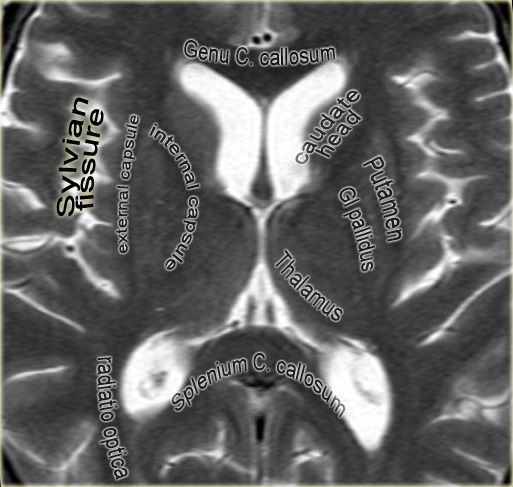
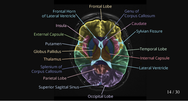
Brain: Atlas of human anatomy with MRI - e-Anatomy - IMAIOS MRI Atlas of the Brain. This page presents a comprehensive series of labeled axial, sagittal and coronal images from a normal human brain magnetic resonance imaging exam. This MRI brain cross-sectional anatomy tool serves as a reference atlas to guide radiologists and researchers in the accurate identification of the brain structures.
MRI anatomy | free MRI axial brain anatomy - Mrimaster.com This MRI brain cross sectional anatomy tool is absolutely free to use. Use the mouse scroll wheel to move the images up and down alternatively use the tiny arrows (>>) on both side of the image to move the images.
Brain MRI Atlas on the App Store Brain MRI Atlas is a FREE app that allows you to navigate through hundreds of of labeled brain structures. It is designed for all healthcare professionals as an interactive study and reference tool. Program Features: - Serial sequential axial T2 FLAIR images of the brain. - Structure labels organized by category.
IMAIOS IMAIOS





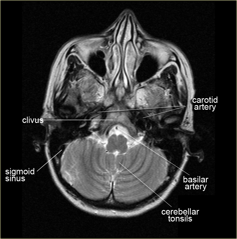
:background_color(FFFFFF):format(jpeg)/images/library/13519/mri-t2-axial-tonsils-cerebellum-level_english.jpg)








Post a Comment for "38 brain mri with labels"