38 human heart with labels and function
Label the heart - Science Learning Hub Label the heart Add to collection In this interactive, you can label parts of the human heart. Drag and drop the text labels onto the boxes next to the diagram. Selecting or hovering over a box will highlight each area in the diagram. Pulmonary vein Right atrium Semilunar valve Left ventricle Vena cava Right ventricle Pulmonary artery Aorta Cross Section of the Heart Diagram & Function | Body Maps The chambers of the heart operate as a 'double-pump' system for the body's circulation. In coordination with valves, the chambers work to keep blood flowing in the proper sequence. The chambers on...
› Make-a-Model-of-a-Heart3 Ways to Make a Model of a Heart - wikiHow Oct 17, 2021 · After pumping the heart without a valve, nothing keeps the blood from moving down the straw. This prevents the blood from moving through the heart and into the body! Remember that the balloon neck is your heart's valve. This example shows how 1 chamber of your heart works with its valve. But inside your body, there are 4 chambers and 4 valves!

Human heart with labels and function
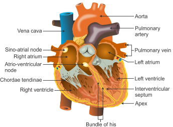
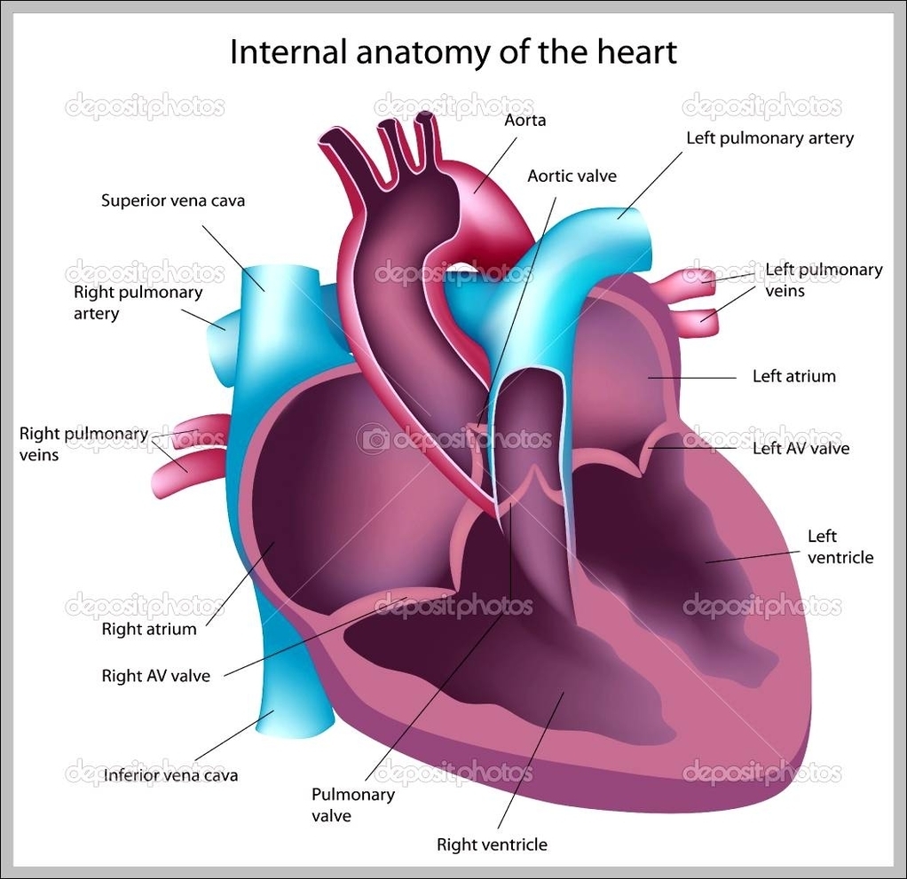
13 parts of the human heart (and its functions ... Parts of the heart and its functions 1. Left atrium 2. Mitral Valve 3. Left Ventricle 4. Aortic sigmoid valve Right atrium 6. Tricuspid valve 7. Right ventricle 8. Pulmonary sigmoid valve 9. Atrial septal defect Interventricular partition 11. The sinus or sinoatrial node 12. Atrioventricular or Aschoff-Tawara nodule 13. Hiscules and Purkinje fibers A Diagram of the Heart and Its Functioning Explained in ... The heart blood flow diagram (flowchart) given below will help you to understand the pathway of blood through the heart.Initial five points denotes impure or deoxygenated blood and the last five points denotes pure or oxygenated blood. 1.Different Parts of the Body ↓ 2.Major Veins ↓ 3.Right Atrium ↓ 4.Right Ventricle ↓ 5.Pulmonary Artery ↓ 6.Lungs PDF Anatomy of Heart Labeled and Unlabeled Images (a) Anterior view of the external heart C' 2019 Pearson Education. Aort'c arch Ligamentum arteriosum Left pulmonary artery Left pulmonary ve ns Auricle of left atrium Circumflex artery Left coronary artery (in atrioventricular sulcus) Great cardiac vein Left ventricle Anterior interventricular artery (in anterior interventricular sulcus) Apex
Human heart with labels and function. Heart Diagram - 15+ Free Printable Word, Excel, EPS, PSD ... Teachers and students use the heart diagram, in biological science, to study the structure and functions of a human being's heart. Friends and colleagues on the other hand may find this diagram template useful when it comes to sending special, personalized gifts to their family members and significant others. Human Heart Diagram Labeled - Science Trends The heart's primary function is to supply the tissues of the body with oxygen and rid the body of carbon dioxide. The pulmonary circuit and the systemic circuit are the two systems of the body that enable the heart to accomplish this. Deoxygenated blood is oxygenated as leaves through the pulmonary circuit. Structure and Function of the Heart - ScienceGeek.net Structure and Function of the Heart. The Human Heart. Refer to this diagram when answering the questions on the right. ... The ascending vena cava is a major vein that returns deoxygenated blood to the heart from the lower part of the body. In this diagram, it is labeled. Diagram of the human heart Images, Stock Photos & Vectors ... Diagram of the human heart royalty-free images. 14,495 diagram of the human heart stock photos, vectors, and illustrations are available royalty-free. See diagram of the human heart stock video clips. Image type.
byjus.com › biology › human-heartHuman Heart - Anatomy, Functions and Facts about Heart Following are the main functions of the heart: One of the primary functions of the human heart is to pump blood throughout the body. Blood delivers oxygen, hormones, glucose and other components to various parts of the body, including the human heart. The heart also ensures that adequate blood pressure is maintained in the body The Anatomy of the Heart, Its Structures, and Functions The heart is the organ that helps supply blood and oxygen to all parts of the body. It is divided by a partition (or septum) into two halves. The halves are, in turn, divided into four chambers. The heart is situated within the chest cavity and surrounded by a fluid-filled sac called the pericardium. Heart Anatomy: Labeled Diagram, Structures, Function, and ... Let's begin with the chambers of the heart. There are 4 chambers, labeled 1-4 on the diagram below. To help simplify things, we can convert the heart into a square. We will then divide that square into 4 different boxes which will represent the 4 chambers of the heart. › productBioDigital | 3D Human Anatomy Software For the first time see human disease and health conditions in interactive 3D. Over 600 health conditions including heart disease, breast cancer, diabetes and more; Optimized for active learning, 3D technology is proven to be a more engaging and accessible way of communicating complex science
› health › healthy-heart-tips28 Healthy Heart Tips Aug 12, 2020 · Eating a diet rich in omega-3 fatty acids can also help ward off heart disease. Many fish, such as salmon, tuna, sardines, and herring, are rich sources of omega-3 fatty acids. byjus.com › biology › diagram-of-heartHeart Diagram with Labels and Detailed Explanation - BYJUS The human heart is the most crucial organ of the human body. It pumps blood from the heart to different parts of the body and back to the heart. The most common heart attack symptoms or warning signs are chest pain, breathlessness, nausea, sweating etc. The diagram of heart is beneficial for Class 10 and 12 and is frequently asked in the ... Anatomy of the Human Heart - Physiopedia The heart is a muscular organ that serves to collect deoxygenated blood from all parts of the body, carries it to the lung s to be oxygenated and release carbon dioxide. Then, it transports the oxygenated blood from the lungs and distributes it to all the body parts The heart pumps around 7,200 litres of blood in a day throughout the body. Human Heart - Anatomy and Functions | Location and Chambers The human heart is the organ that pumps blood throughout the body via the vessels of the circulatory system, supplying oxygen and nutrients to the tissues and removing carbon dioxide and other wastes. Pumping the blood through the arteries, capillaries, and veins is the major function of the heart. It maintains proper circulation of blood.
V Ling: Collector rough details
The 18 parts of the human heart, and their functions ... The 18 parts of the human heart and how they work 1. Myocardium 2. Endocardium 3. Pericardium 4. Right Auricle 5. Right ventricle 6. Tricuspid valve 7. Pulmonary valve 8. Left Auricle 9. Left ventricle 10. Mitral valve 11. Aortic valve 12. Tendon cords 13. Papillary muscles 14. Sinus node 15. Atrioventricular node 16. Atrioventricular fascicule 17.
Describe the working and function of human heart briefly - CBSE Class 11 - Learn CBSE Forum
A Labeled Diagram of the Human Heart You Really Need to ... The human heart, comprises four chambers: right atrium, left atrium, right ventricle and left ventricle. The two upper chambers are called the left and the right atria, and the two lower chambers are known as the left and the right ventricles. The two atria and ventricles are separated from each other by a muscle wall called 'septum'.

Biology Diagrams,Images,Pictures of Human anatomy and physiology: Coronary Arteries
› en › e-AnatomyNormal chest MDCT with anatomic labels | e-Anatomy - IMAIOS Mar 10, 2022 · Pocket Atlas of Human Anatomy: 5th edition - W. Dauber, Founded by Heinz Fene Anatomical variants and notes from the author about the anatomical labeling of the thorax CT: In the lower lobe of the left lung, there is an inconstant subsuperior pulmonary segment that is seen in approximately 30% of individuals, located between the superior and ...

Biology Diagrams,Images,Pictures of Human anatomy and physiology : Lymphatic System
How the Heart Works - The Heart | NHLBI, NIH The heart is an organ about the size of your fist that pumps blood through your body. It is made up of multiple layers of tissue. Your heart is at the center of your circulatory system. This system is a network of blood vessels, such as arteries, veins, and capillaries, that carries blood to and from all areas of your body.
/lungs_alveoli-57ffa7fe3df78cbc284e162b.jpg)
How To Make a Model of the Lungs
The structure of the heart - Structure and function of the ... The structure of the heart. If you clench your hand into a fist, this is approximately the same size as your heart. It is located in the middle of the chest and slightly towards the left.

15 Ways Wolfram|Alpha Can Help with Your Classes—Wolfram Blog
heart | Structure, Function, Diagram, Anatomy, & Facts ... heart, organ that serves as a pump to circulate the blood. It may be a straight tube, as in spiders and annelid worms, or a somewhat more elaborate structure with one or more receiving chambers (atria) and a main pumping chamber (ventricle), as in mollusks. In fishes the heart is a folded tube, with three or four enlarged areas that correspond to the chambers in the mammalian heart.
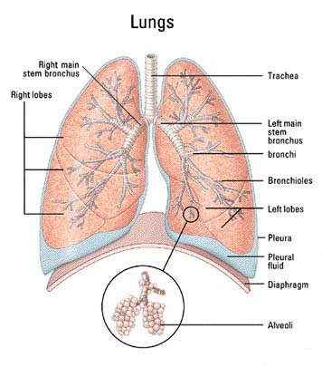
Lungs - microbewiki
Labelling the heart - Science Learning Hub Blood transports oxygen and nutrients to the body. It is also involved in the removal of metabolic wastes. In this interactive, you can label parts of the human heart. Drag and drop the text labels onto the boxes next to the diagram. Selecting or hovering over a box will highlight each area in the diagram.
37 Label The Anatomy Of The Heart - Labels 2021
Heart - Wikipedia The heart is a muscular organ in most animals that pumps blood through the blood vessels of the circulatory system. The pumped blood carries oxygen and nutrients to the body, while carrying metabolic waste such as carbon dioxide to the lungs.

cardiac anatomy and physiology revision :: www.forensicmed.co.uk
Parts Of The Human Heart - Science Trends But few people know all its parts and their essential functions. Broadly speaking, the human heart has one function: to pump blood through the circulatory system all throughout the body, thus supplying both nutrients and oxygen to the body's tissues while also removing all the wastes, including carbon dioxide. The Anatomy of the Human Heart
CLASS NOTES: Basic Heart Anatomy (Vital Signs: Understanding What the Body Is Telling Us)
Heart: Anatomy and Function Your heart is the main organ of your cardiovascular system, a network of blood vessels that pumps blood throughout your body. It also works with other body systems to control your heart rate and blood pressure. Your family history, personal health history and lifestyle all affect how well your heart works. Appointments 800.659.7822

Label Function Human Heart Diagram And Function - Aflam-Neeeak
› en › e-AnatomyAnatomy of the heart and coronary arteries (coronary CT) - IMAIOS Sep 13, 2021 · Anatomy of the human heart and coronaries: how to view anatomical structures. This tool provides access to an MDCT atlas in the 4 usual planes, allowing the user to interactively discover the heart anatomy. The images are labeled, providing an important medical and anatomical tool. The quiz mode makes it possible to evaluate the user's progress.

Circulatory System Diagram | New Health Advisor
Free Heart Worksheets for Human Anatomy Lessons Print out sheet of the human heart with labels - This fun heart worksheet shows kids the different parts of the heart. They'll learn about the left ventricle, the left atrium, the tricuspid valve, and more. Human Heart Clipart - There is a coloring page, heart labeling worksheet and heart anatomy chart. Clipart is a fun way for kids to ...

The Skeletal System-Avery Gillett
Anatomy of a Human Heart - uofmhealth Parts of the human heart . The heart is made up of four chambers: two upper chambers known as the left atrium and right atrium and two lower chambers called the left and right ventricles.. MORE FROM MICHIGAN: Sign up for our weekly newsletter. It is also made up of four valves: the tricuspid, pulmonary, mitral and aortic valves.
.svg/400px-Diagram_of_the_human_heart_(cropped).svg.png)
USMLE Step 1 Review/Cardiovascular - Wikibooks, open books for an open world
Heart (Human Anatomy): Overview, Function & Structure ... The heart is one of the most vital and delicate organs in the body. If it does not function properly, all other organs - including the brain - begin to die from lack of oxygen within just a few minutes. As of 2009, the most common cause of death in the world was heart disease. Most heart disease occurs as a result of age or lifestyle.
Post a Comment for "38 human heart with labels and function"