40 colon diagram with labels
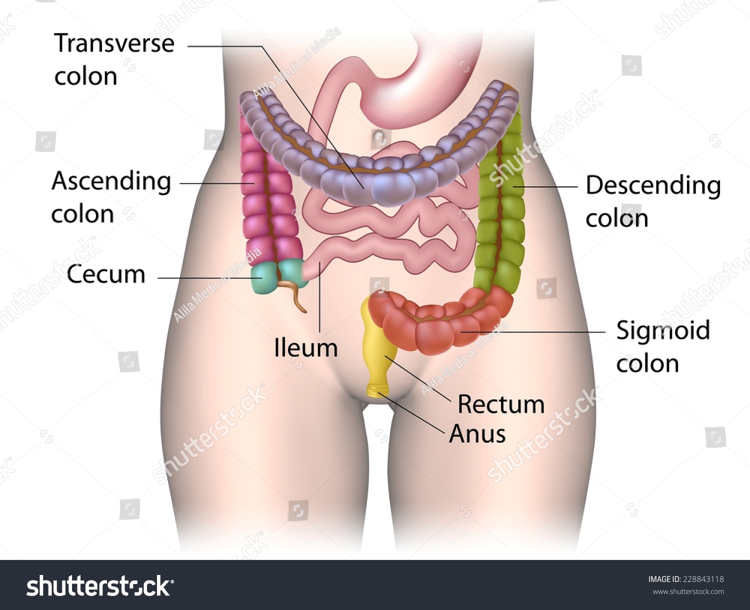
How does the bowel work? Bowel information with diagrams Diagram of the position and sections of the small bowel The colon The colon is divided into 4 sections. Ascending colon The first part of the colon is joined to the small bowel and goes up the right side of the abdomen (tummy). Transverse colon The second section goes across the abdomen from your right to your left side. Descending colon Large intestine diagram labeled - Pinterest We provide you Large intestine structure in easy way also simple Large intestine diagram to draw. And wasy to undersand Large intestine diagram labeled and ...
Sigmoid colon - Definition, Anatomy and Function | Kenhub Sigmoid colon Author: Shahab Shahid MBBS • Reviewer: Jerome Goffin Last reviewed: August 22, 2022 Reading time: 8 minutes The sigmoid colon is part of the hindgut.It is the last part of the colon before the rectum.It acts as a site for water absorption from the faeces, as a site for flatus to be stored before being expelled, and as a site of peristalsis.

Colon diagram with labels
40 Colon diagram Vector Images, Colon diagram Illustrations - Depositphotos 40 Colon diagram Stock Vector Images, Royalty-free Colon diagram Drawings & Illustrations. VectorMine Crohns disease vector illustration. Labeled diagram with diagnosis. VectorMine Ulcerative colitis vector illustration. Labeled anatomical infographic. Anatomy of Colon and Rectum | SEER Training Anatomy of Colon and Rectum. The entire colon is about 5 feet (150 cm) long, and is divided into five major segments. The rectum is the last anatomic segment before the anus.. The ascending and descending colon are supported by peritoneal folds called mesentery.. The right colon consists of the cecum, ascending colon, hepatic flexure and the right half of the transverse colon. Blog - Create a sequence diagram Labeling messages and roles in sequence diagrams Role names for instances of objects/systems and actors are indicated with a colon. E.g. : Customer Conditions (guards) on messages are indicated with square brackets either on the message, or in the frame shape. E.g. [value > $1000] Message numbers can be used to clearly show the sequence of events.
Colon diagram with labels. Label Digestive System Diagram Printout - EnchantedLearning.com Read the definitions below, then label the digestive system anatomy diagram. anus - the opening at the end of the digestive system from which feces (waste) exits the body. appendix - a small sac located on the cecum. ascending colon - the part of the large intestine that run upwards; it is located after the cecum. Liver Diagram with Detailed Illustrations and Clear Labels - BYJUS Liver Diagram. The liver is one of the most important organs in the human body. Anatomically, the liver is a meaty organ that consists of two large sections called the right and the left lobe. The rib cage partly protects the liver and cannot be felt if you were to touch it. However, it can be felt ascending and descending if you were to take a ... Molecular Modeling Database (MMDB) Help Document Range queries are constructed by specifying a lower and upper numerical value separated by a colon (:) to specify the range, followed by a search field name or abbreviation in square brackets, as shown in the examples to the right. You can insert a space on each side of the colon but that is not necessary; the search will work either way. General colon anatomy, with labels Stock Photo - Alamy Download this stock image: General colon anatomy, with labels. - GDP6DM from Alamy's library of millions of high resolution stock photos, illustrations and ...
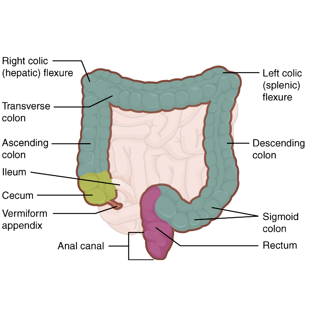
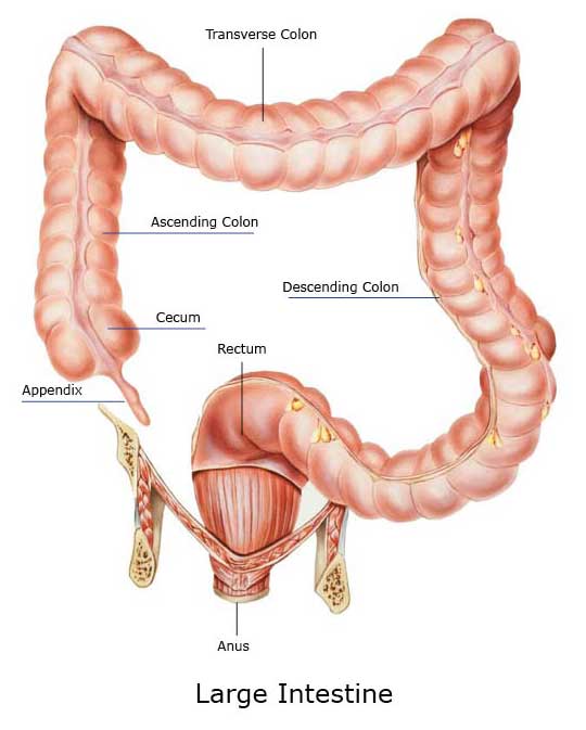
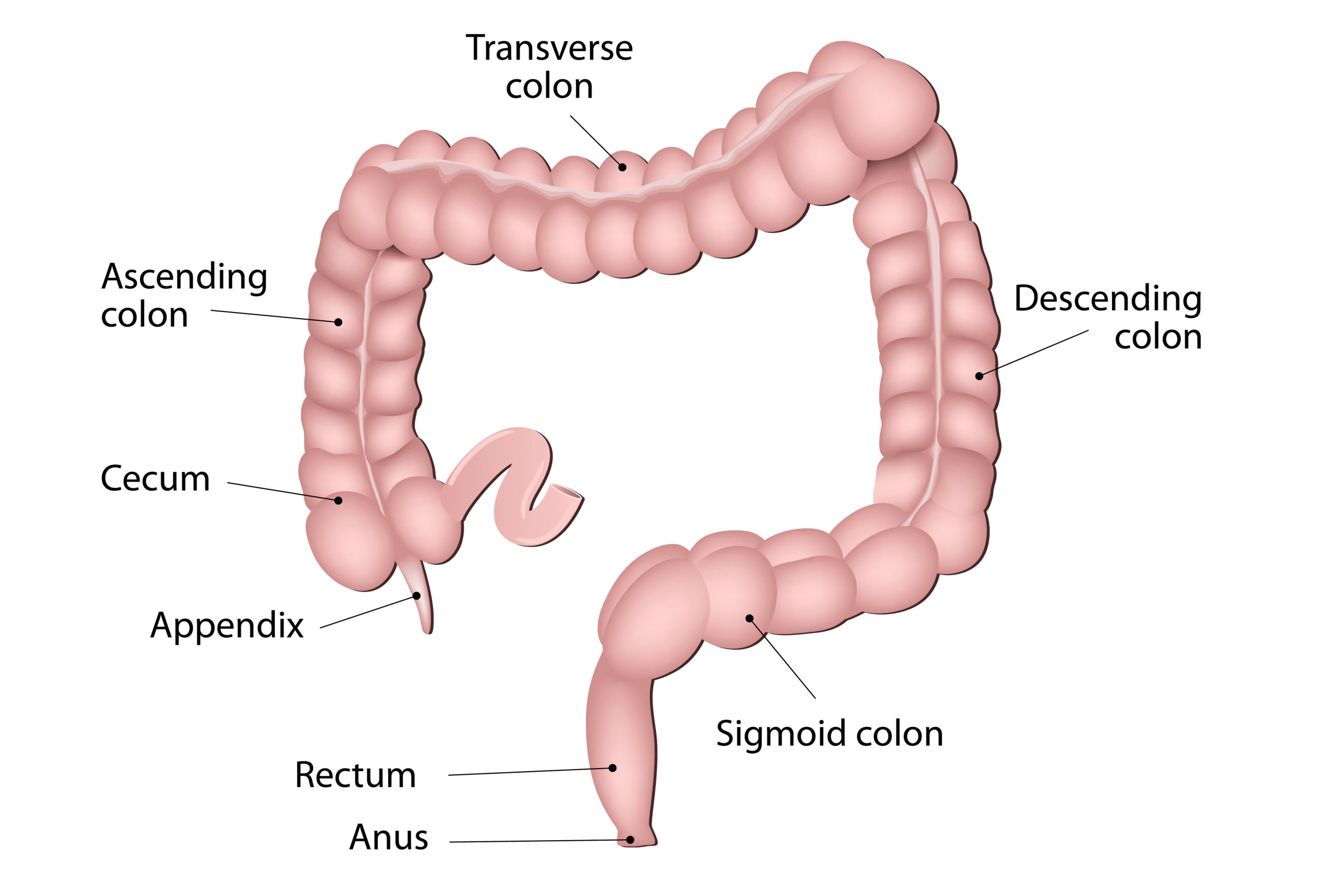
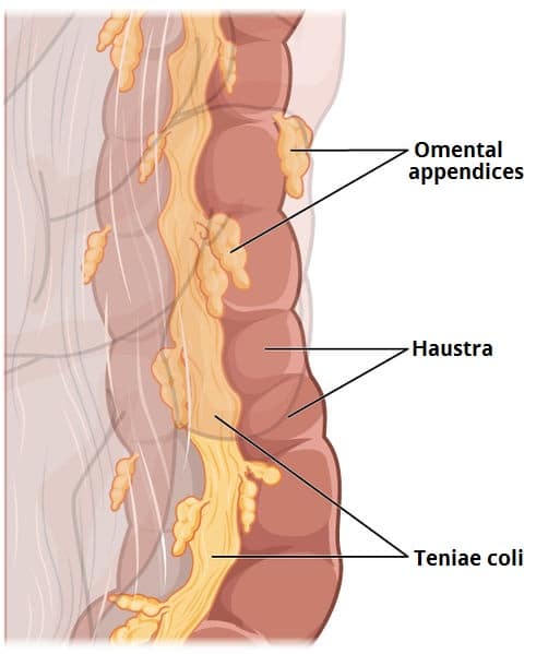
Colon Anatomy - Medical Art Library The colon is divided into four parts: the ascending, transverse, descendingand sigmoid. The ascending and transverse colon meet at the right hepatic flexure(near the liver). The transverse and descending colon meet at the left splenic flexure(near the spleen). The large intestine has three bands of longitudinal muscle fibers called taeniae coli. Colon anatomy: Pictures, features, and function - Medical News Today Click on the body map above to interact with a 3D model of the colon. Cecum and appendix The cecum, pronounced "see-kum," is also called the "proximal right colon." It is a small pouch that... Colon (Large Intestine): Anatomy, Function, Structure - Verywell Health Ascending colon: The ascending colon is the first part of the large intestine.It begins just beyond the cecum (a pouch-like structure at the end the ileum - the part of the small intestine furthest from the stomach) on the bottom right side of the abdomen and ascends (goes upwards) to the area of the abdomen just below the diaphragm. Digestive organs: Diagram, stomach, intestines, and more The large intestine includes the cecum, transverse colon, ascending colon, descending colon, and sigmoid colon. A small, finger-like projection from the large intestine, the appendix, can become...
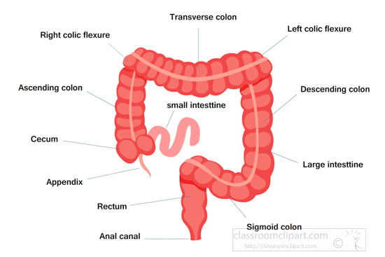
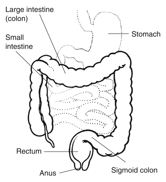
Labeled Diagram of the Human Kidney - Bodytomy The renal medulla comprises a set of 8-18 conical structures called renal pyramids that are surrounded by the cortex. Portions of the cortex between two adjacent pyramids are termed as renal columns. Spread in these pyramids and the cortex, are the functional units callednephrons. The actual filtration of blood occurs in the nephrons. Colon Anatomy (with Small Intestine Label) - National Cancer Institute 720x602. View. Download. Title: Colon Anatomy (with Small Intestine Label) Description: Drawing shows the cecum, ascending colon, transverse colon, descending colon, sigmoid colon, rectum, and anal canal. Also shown is the small intestine. The cecum connects the small intestine to the colon. Course Help Online - Have your academic paper written by a ... Professional academic writers. Our global writing staff includes experienced ENL & ESL academic writers in a variety of disciplines. This lets us find the most appropriate writer for any type of assignment. Large intestine - Wikipedia The large intestine, also known as the large bowel, is the last part of the gastrointestinal tract and of the digestive system in tetrapods.
Large Intestine Anatomy, Parts, Diagram & Major Function ...
Colonoscopy Measurements (cm) from Anal Verge | SEER Training Types of Surgery: Colon; Types of Surgery: Rectum; Radiation Therapy; Commonly Used Drugs; For hands-on exercises, please go to SEER*Educate. Resources. Archived Modules. Updates. Acknowledgements. Colonoscopy Measurements (cm) from Anal Verge. Return to Anatomy of Colon and Rectum. Follow SEER. Contact Information.

Illustration of the large intestine. Taken from Anatomy and ...
The Colon - Ascending - Transverse - Descending - Sigmoid - TeachMeAnatomy The colon (large intestine) is the distal part of the gastrointestinal tract, extending from the cecum to the anal canal. It receives digested food from the small intestine, from which it absorbs water and electrolytes to form faeces. Anatomically, the colon can be divided into four parts - ascending, transverse, descending and sigmoid.
Colon: Anatomy, histology, composition, function | Kenhub
Illustration Picture of Anatomy - Colon - eMedicineHealth The colon is the largest part of the large intestine, extending from the cecum to the rectum. It is 5 feet long and its function is to reabsorb water from digested food and concentrate solid waste material, known as stool. The colon is made of several sections. The ascending colon travels up the right side of the abdomen.
Large intestine: illustration | Radiology Case | Radiopaedia.org
PDF ANATOMIC DRAWINGS OF THE DIGESTIVE SYSTEM Esophageal sphincter Liver ... Colon (C18._).0 Cecum.1 Appendix.2 Ascending.3 Hepatic flex..4 Transverse.5 Splenic flex..6 Descending.7 Sigmoid.8 Overlapping.9 Colon, NOS Yes Yes Yes Yes Yes Yes Yes Yes Yes Yes Yes Yes Yes Yes Yes Yes Yes Yes Yes Yes Yes Yes Yes Yes Yes Yes Yes Yes Yes Yes Yes Yes Yes Yes Yes Yes Yes Yes Yes Yes Yes Yes Yes Yes Yes Yes Yes Yes Yes Yes Yes ...
How does the bowel work? Bowel information with diagrams ...
Simple Guide on Creating Flowchart for Switch Statement Dec 15, 2021 · Each case is then followed by different case labels that always end with a colon (:). The value A, B, and n are case labels that are used for identifying each case individually. Be sure that none of the case labels is the same and each is named according to the preference of execution. For example, two cases have been labeled X.

the large intestine labeling diagram Diagram | Quizlet
Large intestine with labels for the appendix, cecum, ascending colon ... Drawing of the large intestine. The appendix, cecum, ascending colon, transverse colon, descending colon, sigmoid colon, rectum, and anus are labeled.
Colon: Anatomy, histology, composition, function | Kenhub
Large Intestine (Colon) - Diagram - BYJUS Well-labelled Diagram of Large Intestine Large Intestine - Description The large intestine is a 1.5 m long organ that extends from the ileocaecal junction to the anus. It constitutes the cecum with an appendix, ascending colon, transverse colon, descending colon, pelvic or sigmoid colon, rectum and the anal canal.
Colon Diagram Stock Illustrations – 3,341 Colon Diagram Stock ...
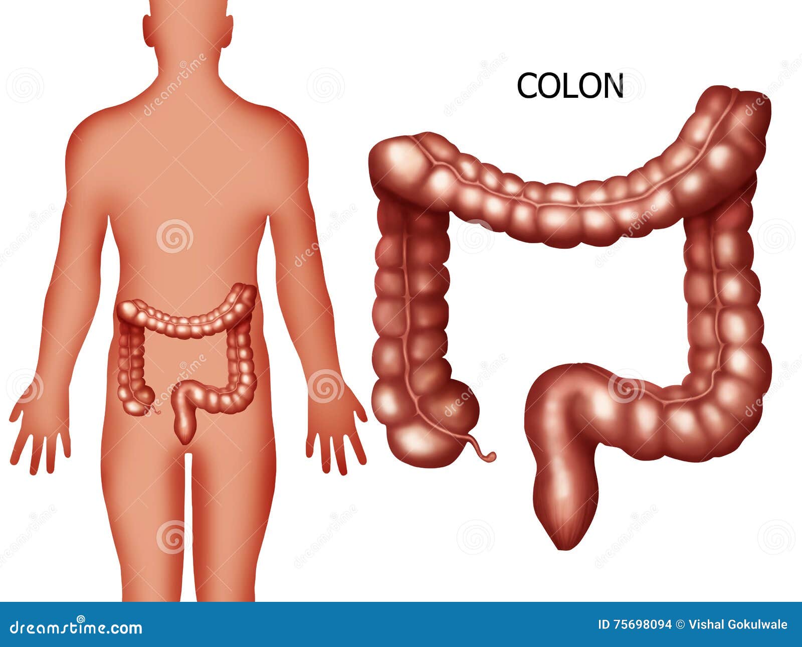
Picture of the Human Colon Anatomy & Common Colon Conditions - WebMD The ileum (last part of the small intestine) connects to the cecum (first part of the colon) in the lower right abdomen. The rest of the colon is divided into four parts: • The ascending colon...
Jejunum's Function in the Small Intestine and Digestive System:
PDF Digestive System Diagram - direzionedidattica-vignola.edu.it Diagram . Large Intestine Mechanical Digestion Chemical Digestion Saliva Hydrochloric Acid Pepsin Trypsin Bile Lipase Stomach Small Intestine Enzymes from Liver and Pancreas Large Intestine (Transverse Colon) Descending Colon System Circulatory Kidneys #1 #2 Water and Vitamins Nutrients The Digestive System Esophagus Mouth On your Digestive ...

Sigmoid colectomy | Alila Medical Images
How to relieve colon pain - zpa.karineedress.shop Sep 06, 2022 · Colon Broom is a plant-based dietary supplement that contains psyllium husk, a fiber that improves the functioning of the digestive system. It also helps with constipation and irregular bowel movements. Colon Broom claims that its products are vegan and gluten-free. The ingredients are carefully researched and safe for human use.

The Large Bowel and Elimination of Faeces - ppt video online ...
Colon Diagram Illustrations & Vectors - Dreamstime Labeled medical diagram with structure and location Colon. Large intestine (colon). Detailed illustration of colon: Ileum, Appendix, Ascending colon, Transverse colon, Descending colon, Sigmoid colon, Rectum and Colon cancer. 3d rendered illustration of a female anatomy with tumor in colon

Normal Colon Anatomy - TrialExhibits Inc.
Colon (Large Intestine): Function, Anatomy & Definition Dec 8, 2021 ... The large intestine includes the colon, rectum and anus. It's all one, long tube that continues from the small intestine and ends at the ...

Human Digestive System With Labels High-Res Vector Graphic ...
Root system - Wikipedia Hasse diagram of E6 root poset with edge labels identifying added simple root position The set of positive roots is naturally ordered by saying that α ≤ β {\displaystyle \alpha \leq \beta } if and only if β − α {\displaystyle \beta -\alpha } is a nonnegative linear combination of simple roots.
The Colon - Ascending - Transverse - Descending - Sigmoid ...
Human Colon Diagram | champion Human Colon Diagram Free Download 2022 by dexter.wehner. Find The BestTemplates at champion. ... Human Teeth Diagram With Labels. Human Teeth Diagram Without Labels. Human Teeth Diagram Canine. Human Teeth Diagram Different Types. Human Heart Diagram Coronary Arteries. Human Resources Dashboard Example.

Foetal large intestine, artwork - Stock Image - C021/2472 ...
2,037 Intestine label Images, Stock Photos & Vectors | Shutterstock Human digestive system, digestive tract or alimentary canal including labels with US spellings. Labelled vector diagram of large intestine.

Anatomy In Motion - The large intestine has a larger width ...
UI Events - W3 Abstract. This specification defines UI Events which extend the DOM Event objects defined in .UI Events are those typically implemented by visual user agents for handling user interaction such as mouse and keyboard input.
Anatomy Clipart - large-intestine-labeled--human-anatomy ...
Human Intestines | Interactive Anatomy Guide - Innerbody Our large intestine consists of 4 major regions: the cecum, colon, rectum, and anal canal. The cecum is a pouch-like dead-end passage that branches inferiorly from the end of the ileum. Fecal matter entering the large intestine from the ileum passes into the cecum before being pushed superiorly into the ascending colon. The appendix is attached ...

unit 5 small and large intestine labeling Diagram | Quizlet
Intestines (Anatomy): Picture, Function, Location, Conditions - WebMD Velvety tissue lines the small intestine, which is divided into the duodenum, jejunum, and ileum. The large intestine (colon or large bowel) is about 5 feet long and about 3 inches in diameter. The...

The Small and Large Intestines | Anatomy and Physiology II
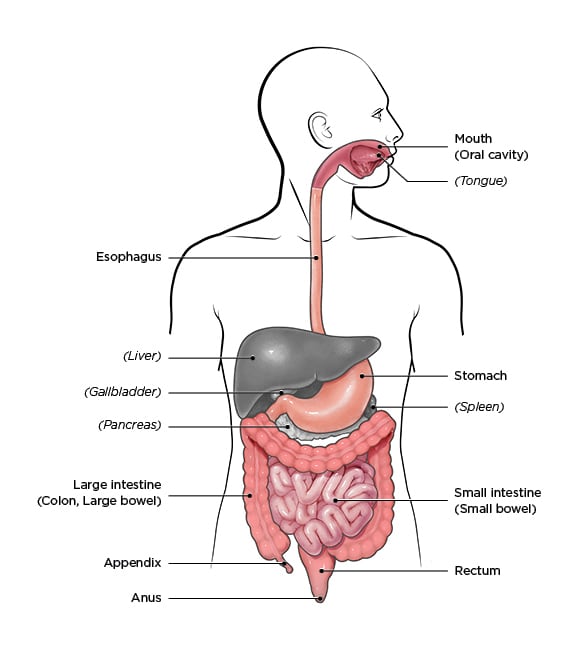
Abdomen and digestive system anatomy: diagrams labeled - IMAIOS Full labeled anatomical diagrams - Anatomy of the abdomen and digestive system: these general diagrams show the digestive system, with the major human anatomical structures labeled (mouth, tongue, oral cavity, teeth, buccal glands, throat, pharynx, oesophagus, stomach, small intestine, large intestine, liver, gall bladder and pancreas).

human digestive system | Description, Parts, & Functions ...
Colon: Anatomy, histology, composition, function | Kenhub The colon forms part of the large intestine and extends between the caecum and the rectum. It is about 1.5 meters in length and consists of four parts: ascending transverse descending sigmoid colon You can recognize it easily through several distinct morphological features like semilunar folds and pouches called haustra.

Labeled medical illustration of the human digestive system ...
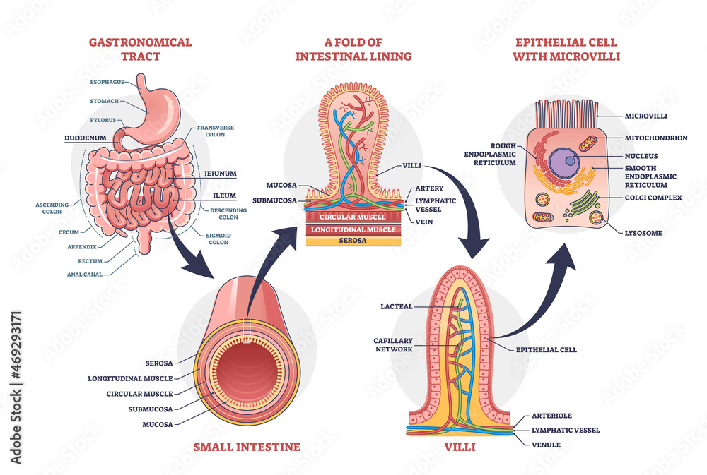
Histology | Colon - The Common Vein The 4 basic layers of the colon: This diagram illustrates the 4 basic layers of the colon. The inner pink layer is the mucosa, the yellow layer beneath the mucosa is called the submucosa, while the red layer is the muscular layer (muscularis) and the 4 th. layer is called the serosa or adventitia.. Courtesy Ashley Davidoff MD

3,289 Colon Diagram Stock Photos, Pictures & Royalty-Free ...
Documentation: DevExtreme - JavaScript Form - DevExpress Specifies whether a colon is displayed at the end of form labels. Applies only to labels outside their editors (see labelMode). showOptionalMark: Specifies whether or not the optional mark is displayed for optional fields. showRequiredMark: Specifies whether or not the required mark is displayed for required fields. showValidationSummary
The Colon - Ascending - Transverse - Descending - Sigmoid ...
Colon Picture Labeling Flashcards | Quizlet Start studying Colon Picture Labeling. Learn vocabulary, terms, and more with flashcards, games, and other study tools. Search. Create. Log in Sign up. Upgrade to remove ads. ... ch.5 pathway of food diagram 14 Terms. olivia_ba. Digestive System Trace 28 Terms. lscott3. RBCs from mitral valve to tricuspid valve->traveling thru big toe 23 Terms ...
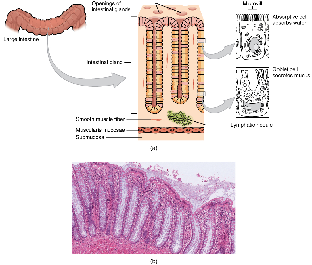
OpenStax AnatPhys fig.23.22 - Histology of the Large ...
Understanding the Human Stomach Anatomy With Labeled Diagrams Given below is a labeled diagram of the stomach to help you understand stomach anatomy. The stomach is divided into four parts. These include: Cardia Fundus Body Pylorus Cardia refers to the section of the stomach that is located around the cardiac orifice. The lower esophageal sphincter lies at the junction where the esophagus meets the stomach.

Functional Anatomy of the Digestive System: Diagram & Organs
Blog - Create a sequence diagram Labeling messages and roles in sequence diagrams Role names for instances of objects/systems and actors are indicated with a colon. E.g. : Customer Conditions (guards) on messages are indicated with square brackets either on the message, or in the frame shape. E.g. [value > $1000] Message numbers can be used to clearly show the sequence of events.

Large intestine structure | Large intestine diagram | Large ...
Anatomy of Colon and Rectum | SEER Training Anatomy of Colon and Rectum. The entire colon is about 5 feet (150 cm) long, and is divided into five major segments. The rectum is the last anatomic segment before the anus.. The ascending and descending colon are supported by peritoneal folds called mesentery.. The right colon consists of the cecum, ascending colon, hepatic flexure and the right half of the transverse colon.
Label Digestive System
40 Colon diagram Vector Images, Colon diagram Illustrations - Depositphotos 40 Colon diagram Stock Vector Images, Royalty-free Colon diagram Drawings & Illustrations. VectorMine Crohns disease vector illustration. Labeled diagram with diagnosis. VectorMine Ulcerative colitis vector illustration. Labeled anatomical infographic.

Colon Picture Image on MedicineNet.com

The Small and Large Intestines | Anatomy and Physiology II
The Lower Digestive Tract with Labels | Media Asset | NIDDK
Small intestine with scientific gastrointestinal tract ...

Colorectal Cancer: Colon and Rectal Cancers : Disabled World
Colon Anatomy (with Small Intestine Label): Image Details ...
The Large Intestine | Complete Anatomy

4,411 Large Intestine Illustrations & Clip Art - iStock
![a: Anatomy of the Large Intestine[6] | Download Scientific ...](https://www.researchgate.net/profile/Asish-Dev/publication/324911391/figure/fig1/AS:622185794260992@1525352061601/a-Anatomy-of-the-Large-Intestine6.png)
a: Anatomy of the Large Intestine[6] | Download Scientific ...
What is inflammatory bowel disease (IBD)? | IBD
Parts Colon Color Coded Labeled Stock Illustration 228843118 ...
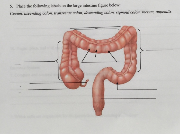
Solved 5. Place the following labels on the large intestine ...
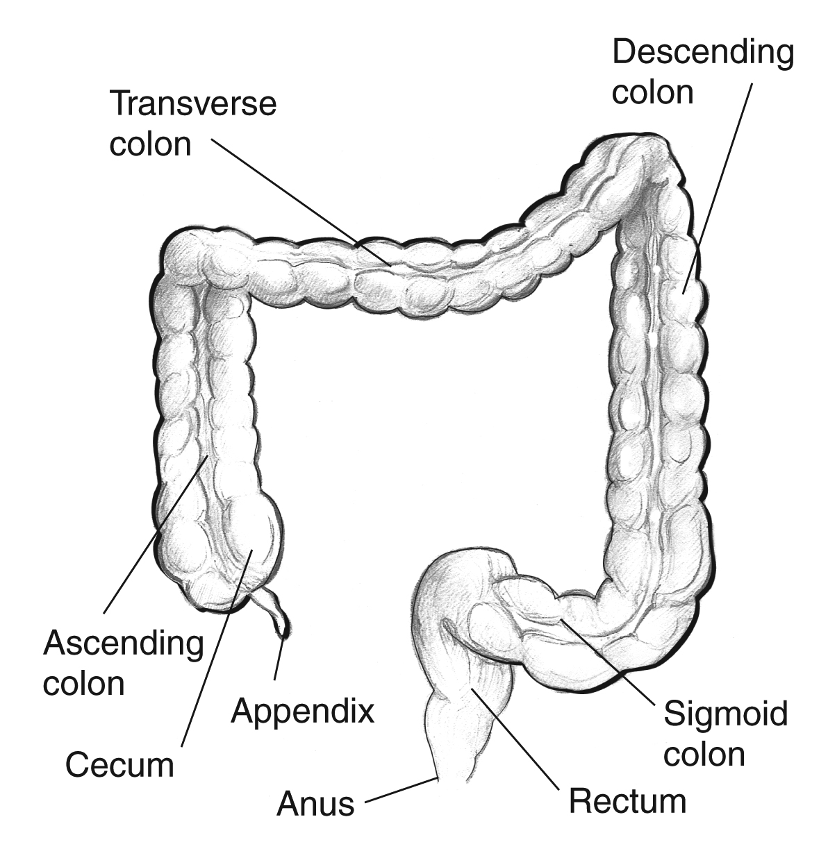
Large intestine with labels for the appendix, cecum ...
Post a Comment for "40 colon diagram with labels"